Tuesday, May 26, 2015

Perch Fish Dissection

Here are some facts about the Perch Fish before we begin the dissection process:
  • They are carnivorous
  • They feed on smaller fish, shellfish, and insect larvae
  • They are found in bodies of water such as ponds or streams
  • They are most commonly found in the Great Lakes
  • Therefore they are a freshwater fish
  • They breathe by using their gills. Gills take in water where it flows through the blood. The blood then proceeds to take out the oxygen from the water.
Fun Fact: There are over 6,000 species in the Percidae family.

External Anatomy
Nares on the front of the fish help in the breathing process.
Gills take in water to filter out the oxygen
the pectoral, pelvic, anal, and dorsal fins help the fish swim
while the caudal fin helps it change direction.  

Internal Anatomy
The heart can be difficult to find at first due to it being
underneath the liver. It pumps blood to the body.
The gills are the breathing mechanism for the fish.


They help with the urinary system by removing waste.
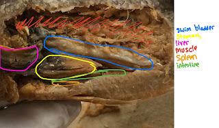
 Swim Bladder-helps the fish stay where it needs to be, no sinking or floating.
Stomach- breaks down ingested food.
Liver-aids in the digestion of food by secreting bile.
Muscle- helps in the movement of limbs.
Spleen- filters the blood.
Intestine- takes nutrients from digested food.


Incision Guide
Cut, in a rectangle, just below the dorsal fin,
right before the anal fin, just above the pelvic fin,
past the pectoral fin.


https://www.youtube.com/watch?v=6jwjqhhxjoU

Crayfish Dissection

Crayfish are found in fresh water like streams, ponds, and ditches. This organism prefers to live in non-polluted water. They can be found under rocks of small streams and creeks. Crayfish eat both plants and animals. They will also eat dead organisms that are relatively fresh. Examples of their diet include worms, insects, insect larvae, and eggs of other organisms. Crayfish are able to live despite hostile conditions due to the wide range of food they are able to consume. Also their ability to consume detritus allows energy transfer of organisms. Crayfish have gills that allows them to retrieve oxygen from water. Crayfish may breathe outside of water if and only if their gills remain moist. In other words, crayfish living in humid environments are able to spend time outside of water. Crayfish can live up to 20-30 years if they hide in holes and do nothing. They reach maturity after 4 years.

Major External Anatomy
Eyes- capture light for vision
Antenna- sensory appendages used to sense food and surroundings
Cheliped- 1st pair of walking legs used for grasping and defense
Walking legs- appendages used for movement
Abdomen- segmented region used to swim backwards
Swimmeret- used for reproduction
Rostrum- protects the sensory appendages of the face
Telson- central swimming paddle
Uropod- small swimming paddles

Green Gland- excretes waste products and balances water in blood
1st-2nd maxilliped- holds food
Mandible- crushes food


We are able to determine the sex of our crayfish by looking at the first set of swimmerets. Males have larger 1st swimmerets to hold the female during mating. They are labeled in blue on the image.

Incision Guide


Begin by cutting along the carapace as shown in the top image. Start from the sides closest to the abdomen and cut towards the head of the cray fish following the curves depicted. Try to cut along the indentations already present on the carapace. Extend the cut closer to the cephalic region. The bottom image shows the cuts made when the crayfish is on its side. Cut from the abdomen towards the cephalic region from the top side first. Then perform the same cut along the bottom side. The image uses highlighted blue markings to demonstrate that this bottom cut should occur without cutting the walking legs.  These legs should be pushed aside to perform this cut.
Major Internal Anatomy
The picture below is kind of blurry. Red highlights are the gills which are used for breathing. The green highlight is the stomach which is used to digest food. The blue highlight is the digestive gland which produces digestive enzymes. The purple is the midgut which is part of the intestines and absorbs nutrients. the yellow is the ventral nerve cord which connects the brain to the rest of the body. The intestines prepare food for secretion. The tail muscles are used for swimming.

Dissection Video

https://www.youtube.com/watch?v=D4gdFtXlvG4


Monday, May 25, 2015

Earthworm Dissection


Earthworms can live in tress, in bark, under rocks, along the river, springs and ponds, they prefer to live in the earth’s rich soil. They get their nutrition from things in the soil such as decaying roots and leaves. Earthworms don’t have lungs, but they breathe through their skin.  

Fun Facts
Earthworms are androgynous, producing both eggs and sperm.

Function of external structures
  • clitellum - produces mucous slime during mating
  • mouth- where the food goes in
  • anus- releases undigested material
  • male genital pore- opening where sperm from that worm is released during mating
  • setae- bristle-like structures that help the worm grip surfaces and to move
  • Prostomium- is a small projection that hangs over the mouth
  • female genital pore- releases eggs
  • sperm groove- Carries sperm from male genital pore to clitellum
  • seminal receptacles- stores sperm received from other worms
  • septum- Divides coelom into compartments
  • segment- contract during movement


Function of internal structures
  • pharynx- sucks in food
  • gizzard - breaks down and grinds the food
  • crop-  stores food
  • esophagus-  food goes through here before it goes to the crop and after the pharynx
  • intestine- food is broken down and absorbed by the blood
  • ventral nerve cord- Relays messages between brain and body
  • ganglia( brian)- masses of tissue containing many nerve cells
  • ventral blood vessel- Transports blood to posterior end of body
  • Dorsal blood vessel- Transports blood to anterior end of body
  • septa- dividing wall between segments
  • nephrida- expel excess water and metabolic waste from the blood
  • testes-produces sperm for fertilization
  • ovaries- where eggs are laid and stored until they mature
  • buccal cavity - Holds for food and opens to Pharynx
  • septum- Divides coelom into compartments
  • seminal receptacles- stores sperm received from other worms
  • central nerve cord-  helps the worm move and feel
  • pseudohearts- pumps blood throughout the body
  • sperm duct- openings in which sperm can pass through
  • oviduct- a tube connecting the ovaries and the uterus
  • typhlosole- provide surface area and increase absorption of nutrients
  • seminal vesicles- storms sperm made by this worm to give away


    Incision Guide-
Cut a slit in the dorsal surface near the posterior pin


 Dissection video


Starfish Dissection


Starfish Dissection

Starfish live in ocean on the sea bottom. Starfish breathe through their feet. Their feet are made of thin tissue that gases can easily move through. Starfish’s tubed feet and tiny bumps all over their bodies transport the oxygen from the seawater to their tissues. Starfish mainly eat clams, oysters’ sand dollars and mussels.

Fun FACT

A starfish can regenerate lost arms. This is useful if the sea star is threatened by a predator. Starfish house most of their vital organs in their arms, so some can even regenerate an entirely new sea star from just one arm and a portion of the star’s central disc. It takes almost a year for this to happen


function of external structures 
 ring of oral spines- spines around the mouth
Central dics- holds all the arms of the starfish 
arms- help with movement and capture prey
Ambulacral groove-contains the tube feet on the oral side and used to pry open the shells of bivalves
  tube feet-  used for feeding, movement, and a little for respiration
eyespot- Senses Light And Dark
madreporite- where water enters in the water vascular system
Spines-  helps protects the starfish
mouth-stomach that is pushed out to eat food


function of internal structures
pyloric ceeca-secretes enzymes for digestion and absorption of nutrients
gonads-  make sperm or eggs
ampulla -Fill up with water and then release to tube feet
ossicles - Plates That Fuse To Form Skeleton
stone canal- a short canal that connects the madreporite to the ring canal around the mouth
ring canal- Circular canal in the center disk
lateral canal-  Branches off of radial canal all the way down the arm and leads into the ampulla


Incision Guide


Dissection video


Grasshopper Dissection

Facts

  • Grasshoppers live on all continents besides Antarctica. Most live in dry environments with lots of grass.
  • They eat leaves, flowers, stems, and seeds. Occasionally they scavenge dead insects.
  • They exchange oxygen and carbon dioxide between their tissues and the atmosphere through air-filled tubes called tracheae. Tracheae open to the environment through small holes called spiracles.
  • In Africa and Asia, grasshoppers are known to be crop pests.

External Anatomy:

  • head, thorax, abdomen: top, middle, bottom regions of grasshopper
  • tympanic membrane: transmit sound from the air to the organism
  • spiracle: hole used by grasshoppers for respiration; air is taken in through them and filtered by hairs in tracheae.
  • ovipositor: tube that delivers eggs
  • antenna: odours, touch, humidity, vibration, wind velocity and direction.
  • compound eyes: can see shape, colour, movement, and tell the distance.
  • mouthpieces: cut and grind food
  • trochanter: attachment point for muscles
  • femur: supports weight of body
  • tarsus: forms ankle joint
  • tibia: connects femur to tarsal segments


Internal Anatomy:
  • heart (not pictured): pumps the fluid to the head from where it filters past the tissues and organs on its way back to the abdomen.
  • crop: stores food
  • gastric caecae: pockets of the stomach that secrete enzymes that break down food
  • intestine: transports waste through the digestive system
  • rectum: passageway for digested waste from the intestine to the anus

Incision Guide:
  • for internal organs

  • for appendages
Dissection Video:


Clam Dissection

Facts

  • Clams live in both freshwater and marine environments.
  • They eat plankton.
  • They take in oxygen through the surrounding water to breathe.
  • “Clams do not have any sense. They cannot smell, hear or even see because they have no noses, ears and eyes.”

External Anatomy:
  • umbo: highest, most prominent part of the shell
  • growth rings: age markers
  • hinge ligament: located near umbo, hinges valves together


Internal Anatomy:
  • gonad: reproductive organ, produces egg or sperm
  • gills:respiratory structures responsible for obtaining water and food
  • digestive gland: used for digestion after passing mouth
  • foot: used to burrow into mud and sand
  • mantle: tissue that lines valve and covers soft body of clam
  • posterior/anterior adductors:on the sides of the clams, used to close shell.
  • exhalant siphon:expels water and waste out of the clam
  • inhalant siphon:brings in oxygen, food, and water into the clam
  • teeth: ridges near anterior end of hinge that interlock when shell is closed


Incision Guide:

  • cut along ventral side
Dissection video:


Sunday, May 24, 2015

Frog Dissection

Frogs are amphibians. This means that they are born in water, but are able to live near and away from water once they become adults. Most frogs are found in areas with warm temperatures. Frogs eat small invertebrates, but some frogs species can be omnivorous. Small frogs eat insects. In the water, frogs breathe through their skin which is a thin membranous tissue that allows gas exchange to occur. Frogs can also breathe through their nostrils using their lungs. Since frogs don't have diaphragms, they must use the bottom of their mouths to push air in and out of the lungs. At rest, frogs use a respiratory surface in their mouth to breathe. Large declines in frog populations have occurred in the past 50 years due to habitat destruction, chemical pollution, climate changes, and many other human activities.
Fun Facts
Frogs’ long legs allow them to leap more than 20 times their body length.
Vocal cords first developed in frogs. Males have vocal cords that fill up with air.

Incision Guide


External Anatomy 
External Nares allow air to enter.
Nictitating Membrane protects the eye.
The tympanum is the eardrum.


Vomerine and Maxillary teeth used to hold prey.
Internal nostrils used for respiration.
Eustachian Tubes provide pressure in the inner ear.
Tongue used to capture prey.
Gullet Opening is the beginning of the digestive tract.

Internal Anatomy
When we opened our frog, we didn't find eggs scattered throughout the internal cavity. However, our frog is a female because we were able to locate the oviducts which produce eggs in female frogs. In male frogs, this structure may be present but it has no vital function (vestigial structure). 


 Function of structures:
Fat Bodies- Like humans, adipose tissue insulates internal organs and protects them.
Heart- Pumps blood to the entire body.
Lungs- Used for respiration and gas exchange.
Peritoneum- Covers internal organs and holds them in place.
Oviducts- Produce eggs in females. Vestigial structure in males.
Liver- Secretes bile and helps digest food.
Stomach- Storage for food. Food digested.
Small Intestine- Most digestion and absorption of nutrients occurs here. Enzyme from the pancreas enter the duodenum.
Large Intestine- Absorbs most fluids. Prepares food for secretion.


Sunday, March 15, 2015

PGlo Transformation


Purpose
The purpose of this experiment was to test the idea that cells can take in genetic information. By using the glo gene, and putting these cells in different situations we were able to see what it takes for a cell to incorporate foreign DNA into their own DNA. This also helped us learn how viruses are able to take over an organism.  

Introduction 
In this experiment we did genetic transformation. Genes contain pieces of DNA that provides the information to make the proteins. Proteins gives an organism their trait. When changed caused by genes it is consider genetic transformation and many use in science. One application would be biotechnology in the fields of agriculture and medicine. For example, with bacteria by moving genes with the help of the plasmid, which the circular DNA. Plasmid DNA usually contains more than one trait that helps the survival of bacteria. Bacteria can transfer plasmids to adapt to new environment. Bacteria becomes resistance to antibiotics because of this transfer. Green fluorescent protein or GFP can by transformed by plasmids. It causes them to glow. When the transformation is done the bacteria will glow fluorescent in the dark. The PGLO plasmid codes for this gene that is resistant to antibiotic ampicillin. To control the rate of proteins that transferred into a cell caused by gene regulation system. Sugar arabinose can be active by GFP. This procedure takes place on antibiotic growth plate

Methods
First, we put in transformation liquid into
each tube; one labeled +pGlo and another
-pGlo.
Then we put them on ice .
Next, we transfered e coli to the tubes. 
We added plasmid DNA only to the
+pGlo tube.
Then we cold shocked the tubes for
10 minutes.
While we waited we labeled our petri dishes
Next we heat shocked the cells by putting
the tubes in a water bath, and back again into
the ice.
Then we added LB nutrient broth to the
mixtures, and mixed.
Lastly, we spreaded the solutions onto
the petri dishes, and let them
incubate for one day. 


Data and Graphs:


Discussion:
The LB/-PGLO plate had the most bacteria that resembled the original untransformed E. coli colonies we initially observed. This makes sense, if we take into consideration the fact that the bacteria were removed from the starter plate, did not have any plasmid added to them, and only had LB (this is broth, or in simpler terms, food) on the plate. Regulating these features makes it a control plate. The other control plate is the LB/amp/-PGLO plate, which had zero growth, because no plasmid was added. Plasmid actually has to be added in order for bacteria to multiply in the presence of ampicillin. The transformation plates include LB/amp/+PGLO and LB/amp/ara/+PGLO. First, both plates had the plasmid for the fluorescent gene added to the E. Coli spread on them. Because of the heat shock we gave the E. Coli, holes could be made in the cell membrane of the prokaryote cells. This means that the PGLO plasmid which expresses the ampicillin resistance gene is incorporated into the E. Coli. Thus the bacteria can survive on the plates that contain ampicillin. On the other hand, cells that were not treated with DNA did not express ampicillin resistance and did not grow on the LB/amp/-PGLO plate. The LB/amp/ara/+PGLO plate had about the same amount of growth as the other transformation plate, but the genes of the plasmid will only be expressed in the presence of arabinose, so it’s on this plate that the bacteria glowed under a UV light. The transformation efficiency (efficiency by which cells take in extracellular DNA and express its genes) for this plate is 406.25 transformants/microgram. This basically reflects how competent prokaryotic cells are at including new DNA. Our results are what they were supposed to be, and they support our belief that only the cells with DNA added to them would be genetically transformed. One way to improve this lab could be to let it go on for more than one day. That way we would try to calculate rate of reproduction in order to grasp how quickly plasmids can be generated in new cells.


Conclusion :
 We found that we were able to transform the DNA of this organism using plasmids. We concluded that E. Coli did not grow. Some bacteria was able to live in the surroundings with ampicillin  and glow under UV light in a environment with arabinose. Based on these  result our  hypothesis came true.